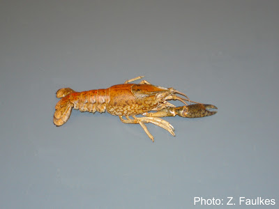
 The start of a year is often a time for growth, renewal, and fresh beginnings, so a picture of a molted exoskeleton seemed particular appropriate.
31 December 2007
Pic of the Moment: 1 January 2008
 The start of a year is often a time for growth, renewal, and fresh beginnings, so a picture of a molted exoskeleton seemed particular appropriate.
24 December 2007
22 December 2007
Marmorkrebs in Wikipedia
 Marmorkrebs have had an article in the German Wikipedia for a while. There is now an entry in the English Wikipedia to go with it. Marmorkrebs are also mentioned in the parthenogenesis article and elsewhere.
Check them out. In true Wiki tradition, feel free to add to them.
18 December 2007
Pic of the Moment: 18 December 2007
Marmorkrebs on the road: SICB 2008
 Your Marmorkrebs.org webmaster will be attending the Society for Integrative and Comparative Biology meeting in San Antonio next month. Although I will not have any Marmorkrebs data on display at the meeting, I will be more than happy to talk with anyone interested in the research prospects for this crustacean. You can meet me at poster P3.73 on Saturday.
11 December 2007
Pic of the moment: 11 December 2007

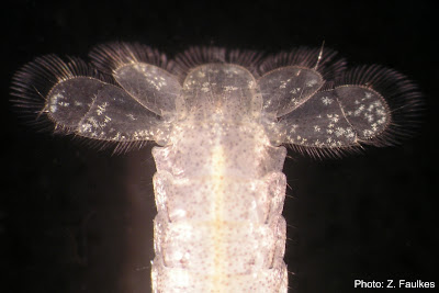
Unlike previous pictures, this picture of a juvenile's tailfan is actually part of a request for information.
What might look to be white spots on the tailfan are seen in close-up (click to enlarge the picture) to be lots of little snowflake shapes. I don't think this is part of the normal juvenile colouration; I strongly suspect this is a fungal infection. The juveniles are growing quite well, so I think that what's happening is that the fungus (if that's what it is) is getting lost each time the animal molts, so nothing serious has come of it yet.
If anyone can confirm that this is a fungus, has suggestions for treatment, and so on, please let me know.
06 December 2007
Sintoni and colleagues, 2007
 Sintoni S, Fabritius-Vilpoux K & Harzsch S. 2007. The Engrailed-expressing secondary head spots in the embryonic crayfish brain: examples for a group of homologous neurons in Crustacea and Hexapoda? Development Genes and Evolution: 217(11-12): 791-799.
http://dx.doi.org/10.1007/s00427-007-0189-5
Abstract
Hexapoda have been traditionally seen as the closest relatives of the Myriapoda (Tracheata hypothesis) but molecular studies have challenged this hypothesis and rather have suggested a close relationship of hexapods and crustaceans (Tetraconata hypothesis). In this new debate, data on the structure and development of the arthropod nervous system contribute important new data (“neurophylogeny”). Neurophylogenetic studies have already provided several examples for individually identifiably neurons in the ventral nerve cord that are homologous between insects and crustaceans. In the present report, we have analysed the emergence of Engrailed-expressing cells in the embryonic brain of a parthenogenetic crayfish, the marbled crayfish (Marmorkrebs), and have compared our findings to the pattern previously reported from insects. Our data suggest that a group of six Engrailed-expressing neurons in the optic anlagen, the so-called secondary head spot cells can be homologised between crayfish and the grasshopper. In the grasshopper, these cells are supposed to be involved in establishing the primary axon scaffold of the brain. Our data provide the first example for a cluster of brain neurons that can be homologised between insects and crustaceans and show that even at the level of certain cell groups, brain structures are evolutionary conserved in these two groups.
Keywords: arthropod • neurophylogeny• evolution • Engrailed • Tetraconata
04 December 2007
Subscribe to:
Comments (Atom)


